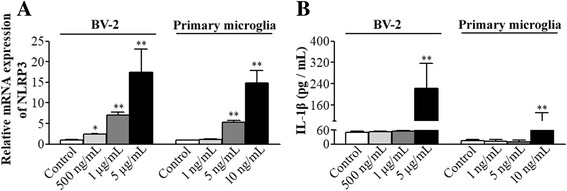
Fig. 3.

NLRP3 priming was induced by LPS stimulation. Different doses of LPS were added to the cell culture media of BV-2 cells or primary microglial cultures for 30 min. a The mRNA of NLRP3 was quantified by real-time qPCR immediately after LPS stimulation. Values are expressed as fold changes over the mean values of blank control. b IL-1β concentration in the supernatant was measured 6 h later. All results are presented as mean ± SD (n ≥ 3). *P < 0.05 and **P < 0.01 compared with the corresponding data of blank control
