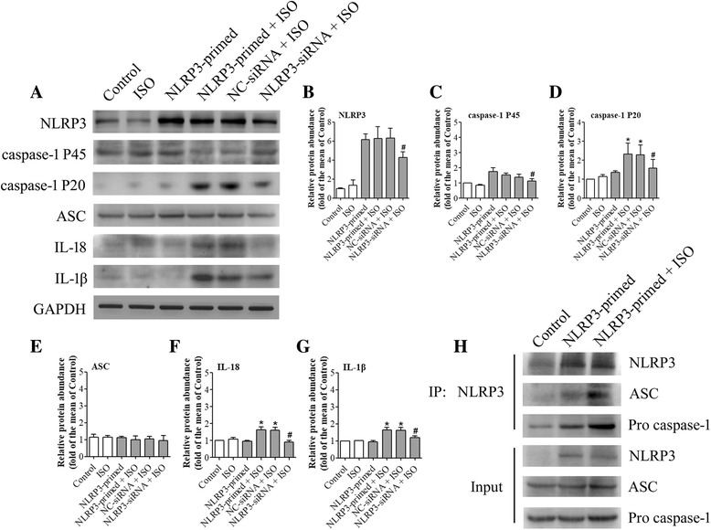
Fig. 6.

NLRP3 knock-down reduced isoflurane-induced NLRP3 inflammasome activation. Cells were transfected with NLRP3 siRNA or negative control siRNA 24 h before NLRP3 priming and exposure to 4% isoflurane for 6 h. NC-siRNA + ISO = negative control of siRNA before NLRP3 priming and isoflurane exposure; NLRP3-siRNA + ISO = NLRP3 siRNA before NLRP3 priming and isoflurane exposure. a Western blot images of NLRP3, caspase-1 P45, caspase-1 P20, ASC, IL-18, and IL-1β. b–g The graphic presentation of each protein abundance of Fig. 6a. h Whole cell protein samples from BV-2 cells were harvested immediately after isoflurane treatment. Immunoprecipitation was performed by using an anti-NLRP3 antibody and was immunoblotted for NLRP3, ASC, and pro-caspase-1. Values are expressed as fold changes over the mean values of NLRP3-primed cells and are presented as mean ± SD (n = 6). *P < 0.05 compared with the corresponding data of group control. #P < 0.05 compared with the corresponding data of NLRP3-primed + ISO
