
Fig. 6.
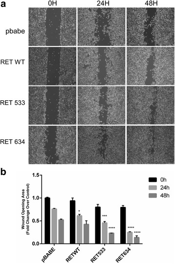
Effect of RET-G533C on cell migration. a. Wound healing assay of HEK293 cells expressing different RET mutants or empty pBABE vector. Cells were scratch-wounded with a micropipette tip (200 μl). Images of cellular migration were taken at times 0 h, 24 h and 48 h using the Leica DFC420 C and Leica Application Suite Software. Magnification, 10X. b. Wound healing was quantified by the ImageJ 64 software using the area of the wound of cells expressing empty pBabe vector at T0 as reference value. Final results represent mean ± SD of three independent experiments and are indicated as fold change over the control in terms of reduction of the wound. Statistical significance was evaluated by Two-Way ANOVA (with multiple comparison Tukey’s test) confronting at each time point the different conditions and indicating the difference over the control (***p < 0.001; ****p < 0.0001)