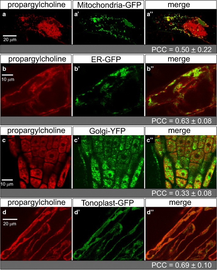
Fig. 6.

Dual visualization of choline phospholipids and GFP marker lines. Arabidopsis seeds were germinated and seedlings were harvested for click chemistry after 7 days on agar media containing 200 µM propargylcholine. a–d Alexa Fluor 594 azide (red) indicates propargyl-PC, a′–d′ GFP (or YFP) (green) marks a specified intracellular compartment, and a″–d″ merged images indicate overlapping expression as yellow or orange, as imaged by confocal laser scanning microscopy. a Hypocotyl epidermal cell shows localization of a mitochondrial marker in comparison to propargyl-PC; b–d root epidermal cells indicate localization of markers for b endoplasmic reticulum (ER), c Golgi apparatus, and d tonoplast. Co-localization was quantified using the Pearson correlation coefficient (PCC) (see Additional file 3: Table S1)
