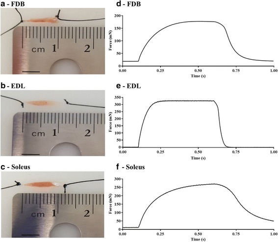
Fig. 3.

Comparison of FDB, EDL, and soleus muscle length, and in vitro isometric contraction profile. Example images of A FDB, B EDL, and C soleus muscle after the tendons were secured with silk suture for subsequent isometric force assessment. Example tetanic (100 Hz) tracings for each muscle: D FDB, E EDL, F soleus
