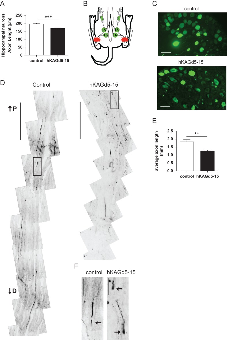
Figure 4.
Overexpression of KIAA0319 decreases axon regeneration in vivo. (A) Quantification of axon length in hippocampal neurons transfected with control plasmid (control; n = 81 neurons) or hKAGd5-15 mutant (n = 76 neurons). (B) Schematic representation of AAV injections at L5 and L6 DRGs (green) and lesion site in the sciatic nerve (red stars). Boxes indicate the region of the sciatic nerve collected for analysis. (C) Representative micrographs of L5 DRGs 10 days after transduction with control AAV-eGFP or hKAGd5-15. Scale bar, 50 μm. (D) Representative micrographs of sciatic nerves from rats with DRGs transduced with either control eGFP (left) or hKAGd5-15 (right) expressing viruses. The micrographs presented were taken 300 μm distally of distance from lesion border. Scale bar, 500 μm. P, proximal; D, distal. (E) Quantification of the length of regenerating axons (considering as origin the distal border of the lesion site) of AAV-transduced DRGs with either control eGFP (n = 5 animals) or hKAGd5-15 (n = 6 animals). (F) Representative growth cones of control and retraction bulbs of hKAGd5-15 transduced DRGs. Axonal tips are highlighted with arrows. Scale bar, 50 μm. Results are expressed in mean ± SEM. **P < 0.01, ***P < 0.001.

