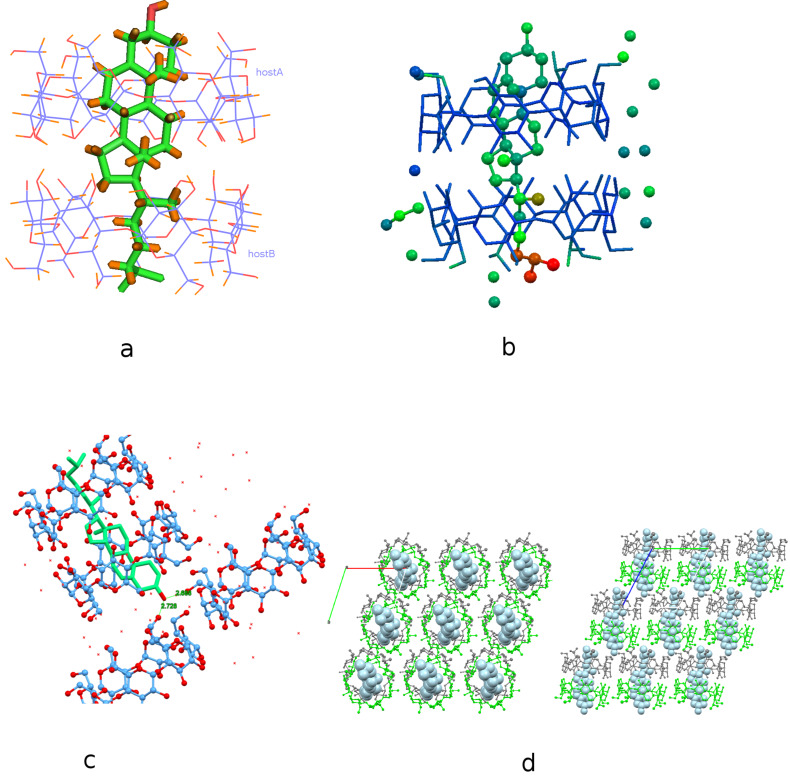
Figure 2.
(a) Crystal structure of the inclusion compound of cholesterol in β-CD dimer. Water molecules are omitted for clarity. (b) The inclusion complex colored by atomic displacement parameters (U’s) using Mercury 3.9. The values of U increase from blue to red colour. (c) The hydroxy group of cholesterol is hydrogen bonded with the hydroxy groups of the primary rim of vicinal β-CD dimers. (d) Inclusion complexes stack along the crystallographic c-axis according to the Intermediate Channel (IM) packing mode. Projection along the c-axis (left) and a-axis (right).

