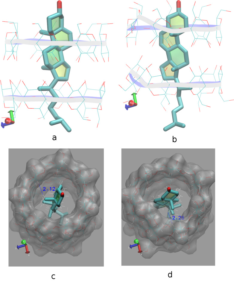
Figure 4.
Representative snapshots of the CHL/β-CD inclusion complex at 0 (a, c) and 11 ns (b, d) in timescale and at 300 K. Water molecules are omitted for clarity. (a, b) Shift of the sterol group of the CHL molecule towards the β-CD dimeric interface. (c, d) H–H interactions between CHL ring A and H5 (initially) or H3 (subsequently) of host A are retained although CHL rotates about hosts’ seven-fold molecular axes. Image rendering was obtained with the VMD visualization program [36].

