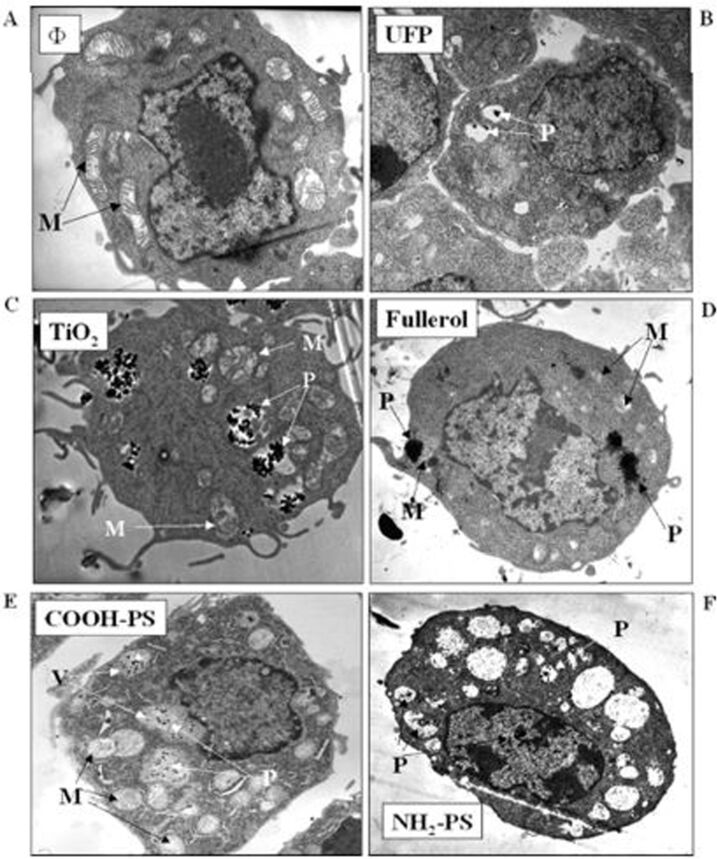
Figure 9.
Electron microscope images show how NPs can penetrate and relocate to various sites inside a phagocytic cell line. (A) Untreated phagocytic cell line (RAW 264.7). Cells were treated with (B) ultrafine particles (<100 nm) (C) TiO2, (D) fullerol, (E) COOH–polystyrene nanospheres, and (F) NH2polystyrene nanospheres. NP exposure was conducted by treating the cells with 10 μg/mL NPs (<100 nm) for 16 h. Labels: M = mitochondria, P = particles [288], copyright 1969, Americal Chemical Society.

