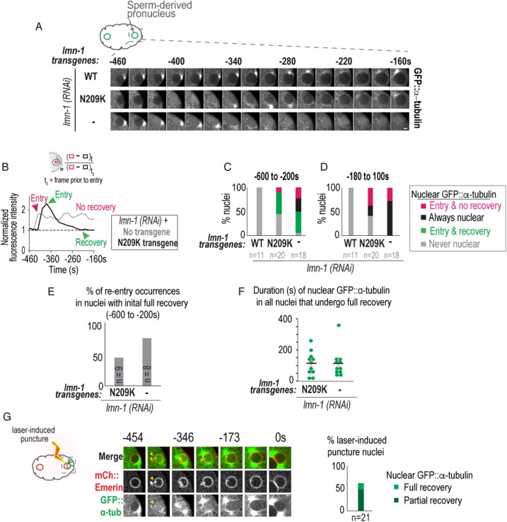
FIGURE 2:
Lamin prevents transient and persistent nuclear ruptures during pronuclear positioning. (A) Representative confocal images of a sperm-derived pronucleus from time-lapse series of indicated conditions. Scale bar, 2.5 μm. (B) (Top) Schematic of method used to measure nuclear GFP::α-tubulin fluorescence intensity levels. (Bottom) Plot of normalized nuclear GFP::α-tubulin fluorescence intensity measurements of indicated nuclei in A. (C, D) Plots of percentage of nuclei during indicated time periods relative to PC regression. (E) Percentage of GFP::α-tubulin nuclear reentry occurrences in nuclei that fully recovered from initial nuclear entry event (green bar in part C). Also see Supplemental Figure S2, A–C. (F) Plot of duration from initial entry to full recovery of nuclear GFP::α-tubulin under indicated conditions. Individual times (green) and mean time ± SEM (black) in seconds are shown. Mann–Whitney test, p = 0.71 n.s. (G) Select images from time series of laser-induced puncture and recovery of sperm-derived pronucleus. The dimensions of this laser-induced puncture are 4 × 2 × 0.5 μm. Arrow points to site of nuclear envelope puncture. Scale bar, 2.5 μm. Time in seconds relative to PC regression. Plot (Right) of percentage of sperm-derived nuclei that fully recover from nuclear GFP::α-tubulin levels (light green) or reach 50% of the nuclear GFP::α-tubulin levels at t0 (partial recovery, dark green). Plot combines data sets of nuclei cut with two different laser punctures sizes (dimensions: 4 × 2 × 0.5 μm [large cut] and 4 × 1 × 0.5 μm [small cut]). n = number of embryos. See also Supplemental Figure S2 and Supplemental Movies 1–3.

