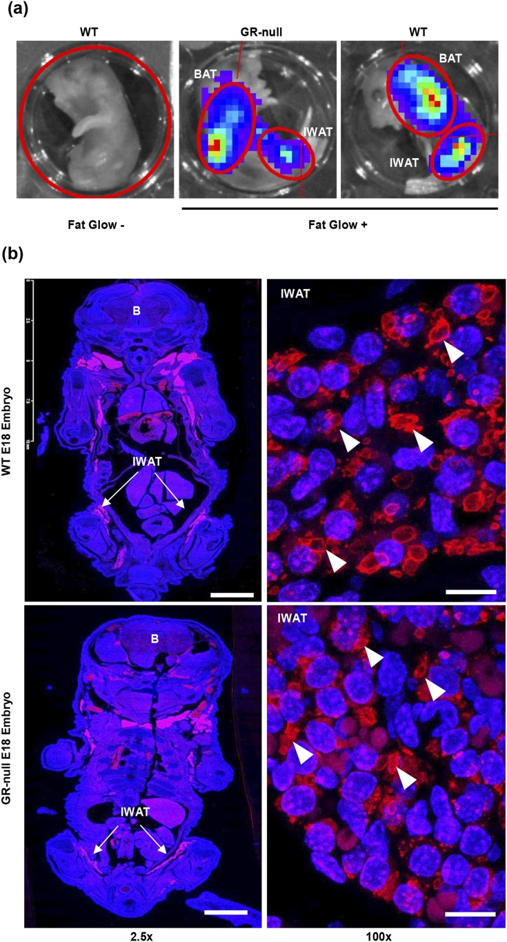
Figure 7.
Early fat pads were present in E18 embryos, independent of GR-status. (a) Bioluminescence was measured in WT and GR-null E18 fat-glow embryos after luciferin injection in the IVIS imaging system. In this model, cre-mediated excision of a floxed STOP cassette upstream of a luciferase reporter was targeted to the adipocyte by selective expression under the adiponectin promoter. Fat-glow‒negative embryos served as a negative control. (b) Coronal sections of fixed and paraffin-embedded WT and GR-null E18 embryos were stained for perilipin (red) to assess for the presence of early adipose depots. Representative images are shown at ×2.5 (scale bar, 2.5 mm) and ×100 (scale bar, 10 µm) power. Nuclei are stained with 4′,6-diamidino-2-phenylindole (blue). Arrowheads represent perilipin-positive lipid droplets. White arrows represent IWAT. B, brain.

