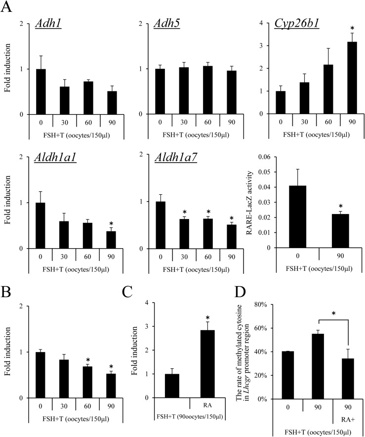
Figure 5.
The negative effects of oocytes on the induction of Lhcgr expression occur via the regulation of methylation in the Lhcgr promoter region in granulosa cells. (A) The effect of oocytes on the expression of Adh1, Adh5, Aldh1a1, Alhd1a7, and Cyp26b1 in granulosa cells. Granulosa cells were collected from ovaries of immature mice after treatment with eCG for 6 hours and were cultured in the medium supplemented with FSH plus T in the presence of 1% FCS. GV-stage oocytes were collected from COCs. None, 30, 60, or 90 of GV-stage oocytes per 150 μL were cultured with granulosa cells in a 96-well plate. Values are given as mean ± standard error of the mean (SEM) of three replicates. The value of granulosa cells that were cultured without denuded oocytes was set as 1, and the data are presented as fold induction. FSH plus T: Granulosa cells were cultured with 50 ng/mL FSH and 10 ng/mL T for 48 hours. Significant differences were observed compared with those treated with no oocytes (P < 0.05). Activity of LacZ enzyme driven by the RARE promoter in granulosa cells cultured with 90 oocytes per 150 μL: Values are shown for RARE-LacZ activity at 595 nm after the protein samples were incubated for 2 hours at 37°C with chlorophenol red-β-d-galactopyranoside. Values are given as mean ± SEM of more than three replicates. Significant differences were observed compared with those treated with no oocytes (P < 0.05). (B) The effect of oocytes on the expression of Lhcgr in granulosa cells. Granulosa cells were collected from ovaries of immature mice after treatment with eCG for 6 hours, and were cultured in the medium supplemented with FSH plus T in the presence of 1% FCS. GV-stage oocytes were collected from COCs. None, 30, 60, or 90 GV-stage oocytes per 150 μL were cocultured with granulosa cells. Values are given as mean ± SEM of three replicates. The value of granulosa cells at 0 hours cultured without denuded oocytes was set as 1, and the data are presented as fold induction. FSH plus T : Granulosa cells were cultured with 50 ng/mL FSH and 10 ng/mL T for 48 hours. Significant differences were observed compared with cells treated with no oocytes (P < 0.05). (C) The additional treatment with RA rescued the negative effects of denuded oocytes on Lhcgr expression in granulosa cells. RA (1 μM) was added to the 150 μL of medium in which granulosa cells were cultured with 90 GV-stage oocytes. Significant differences were observed as compared with cells treated with 90 oocytes per 150 μL (P < 0.05). (D) The effect of oocytes on the methylation levels of Lhcgr promoter region in granulosa cells. Granulosa cells were collected from ovaries of immature mice, after treating with eCG for 6 hours, and were cultured in the medium supplemented with FSH plus T in the presence of 1% FCS. GV-stage oocytes were collected from COCs. GV-stage oocytes (90 per 150 μL of medium) were cultured with granulosa cells in a 96-well plate. Values are given as mean ± SEM of three replicates. FSH plus T: Granulosa cells were cultured with 50 ng/mL FSH and T10 ng/m T for 48 hours. RA (1 μM) was added to the in vitro culture of granulosa cells with 90 GV-stage oocytes per 150 μL of medium. Significant differences were observed as compared with cells treated with 90 oocytes per 150 μL of medium. *P < 0.05.

