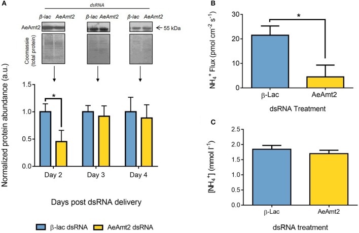
Figure 6.
Effects of AeAmt2 dsRNA treatment on AeAmt2 abundance in the anal papillae, excretion from the anal papillae, and concentration in the hemolymph. (A) Representative Western blots (1 representative of 3 replicates) and densitometric analysis of AeAmt2 (55 kDa) in the anal papillae on days 2–4 following dsRNA treatment. Each group was normalized to Coomassie total protein staining, used as a loading control, and is expressed relative to the β-lac control group (assigned a value of 1, n = 3). Asterisk denotes a significant difference in normalized protein expression compared to the β-lac control group based on an unpaired one-tailed t-test of relative density (p = 0.0491). (B) Scanning ion-selective electrode technique (SIET) measurements of flux across the anal papillae epithelium 2 days post β-lac and AeAmt2 dsRNA treatment (n = 10). Asterisk denotes a significant difference in efflux compared to the β-lac control group based on an unpaired t-test (p = 0.0125). (C) Ion-selective microelectrode measurements of concentration in the hemolymph of larvae at 2 days post β-lac and AeAmt2 dsRNA treatment (n = 10). Data is shown as mean values ± SEM.

