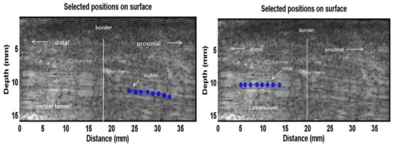
Fig. 4.
A B-mode image of the tendon inside and outside the carpal tunnel in the ex vivo human cadaveric hand testing. 8 locations over a length of 8 mm in the tendon inside the carpal tunnel and 8 locations over a length of 8 mm in the tendon outside the carpal tunnel were selected to measure the tissue motion using ultrasound tracking beams, respectively. The blue dots represent the relative locations of the selected points on the obtained ultrasound B-mode image.

