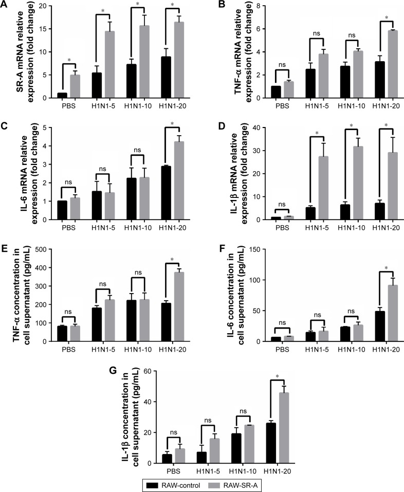
Figure 8.
RT-qPCR and ELISA results of RAW-SR-A and RAW-control cells stimulated by H1N1 influenza.
Notes: RAW264.7 cells were plated as 106 cells/well in a six-well plate and cultured to 70%–80% confluence. After overnight incubation, the culture medium was replaced with serum-free DMEM containing H1N1 influenza virus at an MOI of 5, 10, and 20 for 24 h with 2 μg/mL Trypsin-TPCK and 25 mM HEPES buffer. The supernatant was collected for ELISA analysis and the adherent cells were collected for real-time PCR. (A) Level of SR-A was significantly higher in RAW-SR-A cells, and it increased as cells were treated with higher concentration of H1N1 influenza virus. (B–G) Similar to previous results, mRNA and protein levels of TNF-α, IL-6, and IL-1β were much higher in RAW-SR-A cells. (*p<0.05 vs the control group).
Abbreviations: ELISA, enzyme-linked immunosorbent assay; ns, nonsignificant; RT-qPCR, real-time reverse transcription quantitative polymerase chain reaction.

