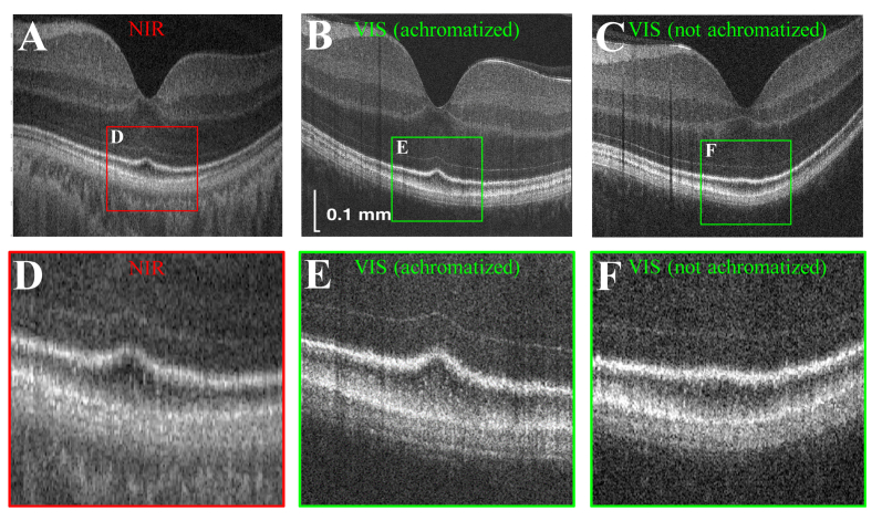
Fig. 3.
Comparison of near-infrared (NIR) light OCT (A,D) and visible (VIS) light OCT, with (B,E) and without (C,F) achromatization in the same eye. Near-infrared (A,D) and visible light OCT images without achromatization (C,F) consist of 2048 axial scans with transverse resolutions of 15 μm and 10 μm respectively. Visible light OCT images with achromatization (B,E) consist of 4096 axial scans with a transverse resolution of 5.2 μm. All images are on a logarithmic scale.

