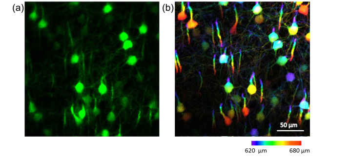
Fig. 4.
In vivo three-photon images of neural cortex in a Thy1-YFP transgenic mouse. (a), projection of 3D volume neurons and neurites in cortex of the awake mouse taken with Bessel-beam at a frame rate of 1 Hz (image averaged from 10 frames afterwards). (b), mean intensity projection of a 65-μm-thick image stack collected at 1-μm z steps in point-scanning mode, from the same region as in (a). The stack covers from 620 μm to 685 μm below the dura, with structures color-coded by depth. The image of each layer was averaged from 3 frames, with a post objective power of 10 mW for Gaussian beam scanning and 110 mW for Bessel beam scanning.

