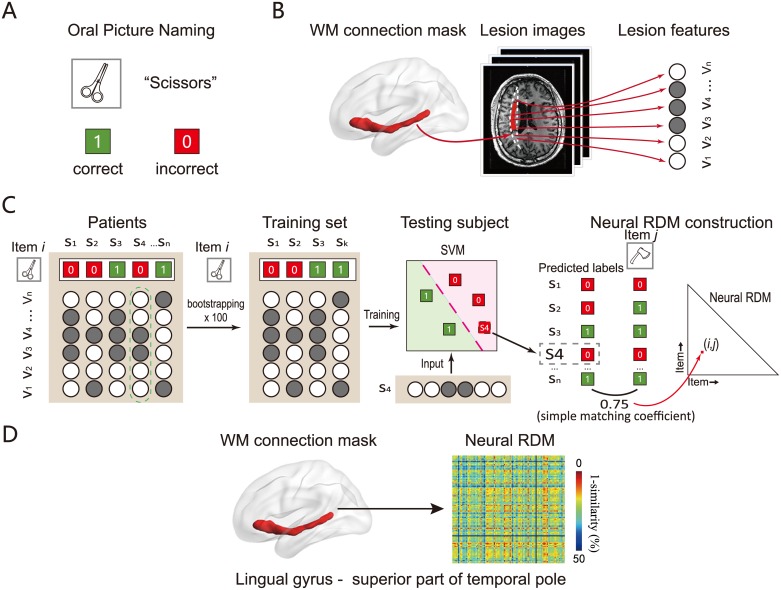
Fig 1. A flowchart for constructing a neural RDM in a WM connection.
(A) The neuropsychological test. We asked patients to complete a picture-naming task containing 100 items. The response for each item was scored as 1 if correct or 0 if wrong. (B) The lesion mask (manually traced in T1 image) in a given patient was converted to MNI space and was then overlapped with the WM connection template constructed from a healthy population [27] to extract the voxel-wise lesion pattern on each WM connection. (C) The SVM classifier was trained on the naming accuracy of one item i (e.g., scissors) and lesion patterns on a WM connection in some patients (see Materials and methods) and then used to generate the predicted naming score in the testing patients (1 or 0). The correspondence (simple matching coefficient) between the predicted score and the actual naming score of each of the other items (e.g., axe) across patients was calculated. This correlation reflects to what degree the lesion features that were useful to predict naming accuracy of item i could also be useful to predict item j, and thus was taken as the neural similarity between the naming process of the training item i and this other item j (scissors–axe similarity) on this connection. All cross-item and within-item similarity could be obtained this way, resulting in a 100 × 100 similarity matrix for this connection. (D) A sample neural RDM of a WM connection (between MTG and superior ATL). The values of dissimilarity were 1-similarity (obtained in C); red indicates low dissimilarity (high similarity) and blue high dissimilarity (low similarity). The object line drawings were done by the first author Y.F.; The brain figure was generated using BrainNet Viewer [28]. ATL, anterior temporal lobe; MTG, middle temporal gyrus; RDM, representational dissimilarity matrix; SVM, support vector machine; WM, white matter.

