
Fig. 13.
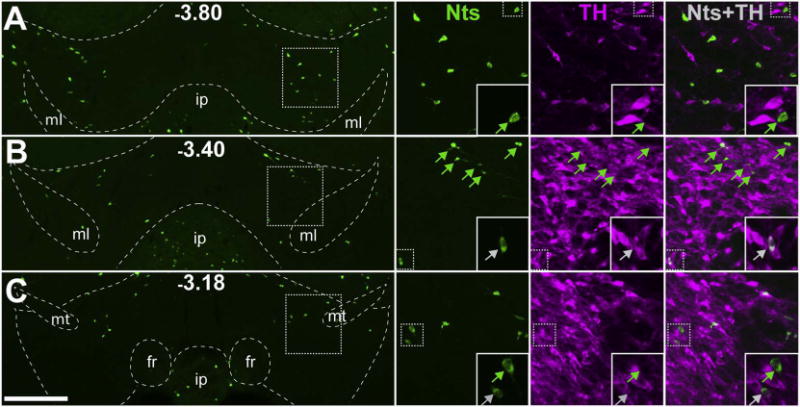
Minimal Nts expression in the VTA. NtsCre;GFP mice (n = 6) were used to determine the number and distribution of VTA Nts neurons and whether or not they co-express TH. A, B, C) Representative images of GFP-identified Nts neurons across three different bregma coordinates in the VTA of NtsCre;GFP mice (scale bar = 200um). Insets highlight individual Nts neurons and the presence or absence of colocalization with TH (greyarrows = TH+ Nts neurons, green arrows = TH- Nts neurons). These data demonstrate that of the few GFP+ cells found in the VTA, the majority do not colocalize with TH and are not DAergic. ml = medial lemniscus, ip = interpeduncular nucleus, fr = fasciculus retroflexus, mt = mammillothalamic tract. (For interpretation of the references to colour in this figure legend, the reader is referred to the web version of this article.)