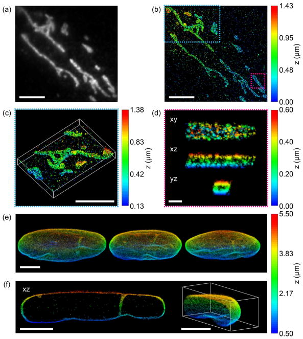
Figure 4.
Performance of TILT3D for 3D single-molecule super-resolution imaging in mammalian cells. (a) 2D diffraction limited image of mitochondria in a HeLa cell immunolabeled with Alexa Fluor 647. (b) 3D super-resolution reconstruction of the sample shown in (a) acquired using the double-helix PSF. (c) 3D view of the mitochondria shown in the blue dashed rectangle in (b). (d) xy, xz, and yz views of a single mitochondrion shown in the magenta rectangle in (b). (e) Three different views of a 3D super-resolution reconstruction of the entire nuclear lamina in a HeLa cell. Here, lamin B1 was immunolabeled with Alexa Fluor 647. Single molecules were detected in several thick, overlapping slices using the double-helix PSF, while fiducial beads were detected using the 6-μm Tetrapod PSF. (f) The xz view shows a 1.3-μm thick slice through the cell in the reconstruction in (e), where lamin meshwork enveloping an intranuclear channel is visible. The right panel shows a cap of the reconstruction in (e). Scale bars are 500 nm in (d) and 5 μm in all other panels. Figure adapted from Ref. 34 with permission.

