
Fig. 1. Overview of SurfaceChIP protocol and profiling of H3K4me3 and H3K27me3 marks in GM12878 cells.
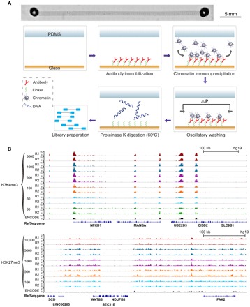
(A) Microscopic image of a single-channel device (stitched from multiple images) and steps involved in SurfaceChIP-seq. The microfluidic channel had dimensions of 40 mm × 1 mm × 60 μm. There were supporting pillars (50 μm in diameter) inside the channel to prevent collapse. (B) Normalized H3K4me3 signals generated using 30 to 5000 cells per assay and H3K27me3 signals using 100 to 10,000 cells per assay. ENCODE data (GSE29611) were included for comparison.