. 2018 Mar 9;7:e30789. doi: 10.7554/eLife.30789

Figure 3. ZHP proteins play a central role in chiasma formation and meiotic chromosome segregation.

(A–D) Upper: DAPI-stained oocyte nuclei at late diakinesis. Each panel shows a representative nucleus. Lower: Graphs indicating the distribution of DAPI-staining bodies observed at diakinesis. ZHPs tagged with AID in a Psun-1::TIR1 background were treated with or without auxin for 24 hr. In the absence of auxin, each nucleus contains six bivalents (homolog pairs held together by chiasmata). Depletion of ZHP-1 or -2 leads to a marked decrease in bivalents per nucleus (8–12 DAPI-staining bodies, or 0–4 bivalents), while depletion of ZHP-3 or -4 results in a complete loss of bivalents (12 DAPI-staining bodies). n is the number of nuclei scored for each condition or genotype. Depletion of ZHP-1,–2, −3 or −4 significantly increase the number of DAPI-staining bodies (***p<0.0001 by Chi-square test for trend). The difference between ZHP-1 depletion and ZHP-2 depletion or ZHP-3 depletion and ZHP-4 depletion is not significant (p=0.7537 and 0.9760, respectively, by Chi-square test for trend). The difference between depletion of ZHP-1 or ZHP-2 and depletion of ZHP-3 or ZHP-4 is significant (***p<0.0001 by Chi-square test for trend). (E) Frequencies of males and viable embryos observed among the whole broods in wild-type, zhp-1, zhp-2 and zhp-3 null mutant hermaphrodites. (F–G) Sex chromosomes and autosomes show a similar reduction in chiasma formation in the absence of ZHP-1. (F) High magnification images of nuclei at late diakinesis from zhp-1 heterozygous controls and null mutant hermaphrodites, hybridized with FISH probes recognizing either the right end of X-chromosome or 5S rDNA on Chromosome V, and stained with DAPI. (G) Graph showing the frequency of bivalents observed for Chromosome V and the X Chromosome. ‘Expected value’ is the frequency for any single chromosome, given the average number of bivalents we observed, and assuming that they were distributed equally among six chromosome pairs. Data were derived from the number of DAPI-staining bodies in zhp-1 heterozygotes and null mutants. Data are represented as mean ±SEM from three independent experiments (n = 103 and 145 nuclei, respectively). Significance was tested using the two-sided Student’s t-test. Scale bars, 5 µm.

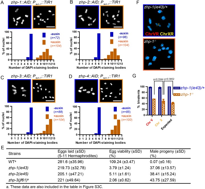
Figure 3.

Figure 3—figure supplement 1. Chiasma formation in zhp-1 or zhp-3 null mutants.

Figure 3—figure supplement 1.

(A–C) Null mutations in zhp-1 and zhp-3 quantitatively recapitulate the effects of AID-mediated depletion. Upper: Representative examples of DAPI-stained oocyte nuclei at diakinesis. Lower: Graphs indicating the distribution of nuclei containing various numbers of DAPI-staining bodies. Note: the observed number of DAPI-staining bodies, on average, is slightly lower than the true mean, since occasionally univalent or bivalent chromosomes in the same nucleus cannot be clearly resolved. (A) Auxin treatment does not impair chiasma formation in wild-type hermaphrodites (p=0.8670 by Chi-square test for trend). (B) A few bivalents are detected in zhp-1 null mutants, as in zhp-1::AID animals treated with 1 mM auxin. (C) No bivalents are observed in zhp-3 null mutants, as in zhp-3::AID hermaphrodites exposed to auxin. The difference in bivalents observed in zhp-1 and zhp-3 null mutants is significant (p<0.0001 by Chi-square test for trend). n is the number of nuclei scored for each condition or genotype. Scale bars, 5 µm.
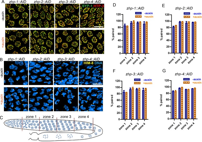
Figure 3—figure supplement 2. ZHP are dispensable for homolog pairing and synapsis.

Figure 3—figure supplement 2.

(A) Projection images of representative pachytene nuclei stained for SYP-1 (green) and HTP-3 (red), revealing normal synapsis in ZHP-depleted worms. Animals expressing the indicated transgene, along with germline-expressed TIR1 protein (Zhang et al., 2015) were incubated on plates with or without 1 mM auxin for 24 hr before dissection. Scale bars, 5 µm. (B) Images of representative early prophase nuclei stained for HIM-8 (yellow), which specifically marks the pairing center region of the X chromosome (Phillips et al., 2005), and DNA (blue). Robust pairing of HIM-8 foci in early meiotic prophase is observed. Homologous pairing at other chromosomal loci was confirmed by FISH (data not shown). Scale bars, 5 µm. (C) Diagram of a hermaphrodite gonad, indicating the four zones in which homolog pairing was scored. (D–G) Quantification of the pairing of X chromosomes. Gonads were divided into four zones of equal length, spanning the meiotic entry through late pachytene region. Pairing was evaluated as previously described (Harper et al., 2011). The average percentage of nuclei with paired X chromosomes is plotted for each zone. Data are presented as mean ±SD (n = 3 gonads for each genotype or condition). No pairing defects were detected following depletion of ZHP-1,–2, −3, or −4 (p=0.7059, 0.3631, 0.6188, 0.3064, 0.0554, 0.5515, 0.2639, 0.4158, 0.1893, 0.9247, 0.8599, 0.3740, 0.7631, 0.1672, 0.7879 and 0.2935, respectively, from zone 1 to zone 4, and zhp-1::AID to zhp-4::AID, two-sided Student t-test).
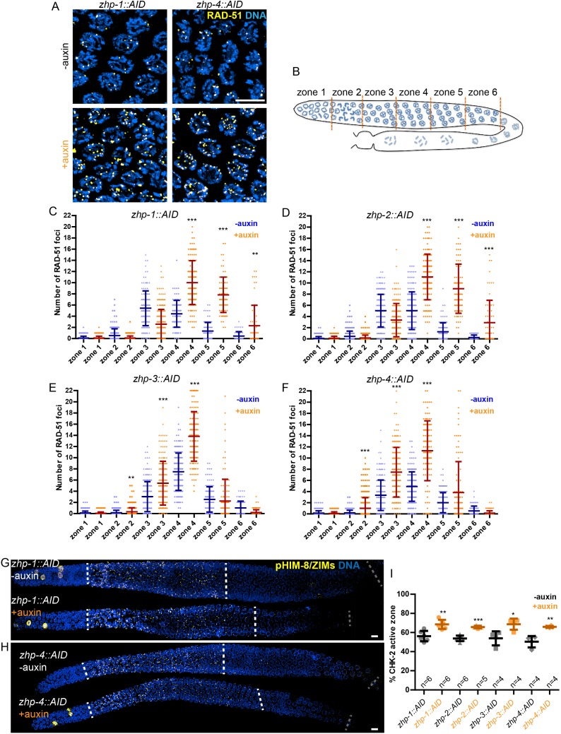
Figure 3—figure supplement 3. ZHPs are dispensable for DSB induction and the crossover assurance checkpoint.

Figure 3—figure supplement 3.

(A) Projection images of mid-pachytene nuclei stained for RAD-51 (yellow) and DNA (blue), showing accumulation of DSB repair intermediates in the absence of ZHP-1 or ZHP-4. Scale bars, 5 µm. (B) Diagram of a hermaphrodite gonad, indicating the six zones in which RAD-51 foci were scored. (C–F) Quantification of RAD-51 foci. Gonads were divided into six zones of equal length, spanning the premeiotic through late pachytene region. The number of RAD-51 foci in each nucleus is presented for each zone. n = 167, 229, 160, 200, 133, 209, 100, 131, 87, 87, 44, 62, 222, 258, 227, 274, 190, 173, 158, 151, 109, 100, 68, 68, 352, 251, 283, 296, 197, 172, 167, 138, 124, 119, 76, 103, 338, 317, 281, 290, 227, 171, 194, 111, 134, 102, 74 and 102 nuclei, respectively, from zone 1 to zone 6, and zhp-1::AID to zhp-4::AID. four gonads were scored for each genotype or condition. **p=0.0084 and 0.0028, respectively, ***p<0.0001, Mann-Whitney test. (G–I) CHK-2 activity is extended in the absence of ZHP proteins. Low magnification images of gonads stained for pHIM-8/pZIMs (yellow) and DNA (blue). The upper gonad in each panel is a non-auxin treated control, while lower panels show gonads from animals depleted of ZHP-1 (G) or ZHP-4 (H) for ~24 hr. Scale bars, 5 µm. (I) Quantification of the ‘CHK-2 active zone,’ defined as the length of the region of pHIM-8/ZIM staining as a fraction of the region from meiotic onset to the end of pachytene before nuclei form a single file. n = number of gonads scored for each genotype or condition. *p=0.0199, **p=0.0018 and 0.0021, respectively, ***p<0.0001, two-sided Student t-test.