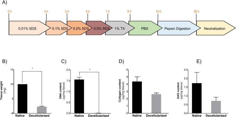
Figure 1. Preparation and characterization of hDLM substrates.
A) Schematic representation of hDLM preparation protocol that consists of decellularization through treatment of liver slices with increasing concentrations of SDS and pepsin digestion followed by neutralization required for gel formation. B) Wet weight of tissue before and after decellularization process shows a 77.1% reduction in tissue mass (p<0.05). C) DNA quantification shows that over 99% of the DNA content is effectively removed from tissue during the decellularization process p<0.05. D) Total collagen content revealed a 40% reduction during decellularization. E) Glycosaminoglycan content analysis showed that 40% of GAGs was preserved in the decellularized tissue. (n = 3, error bars represent Standard Deviation).

