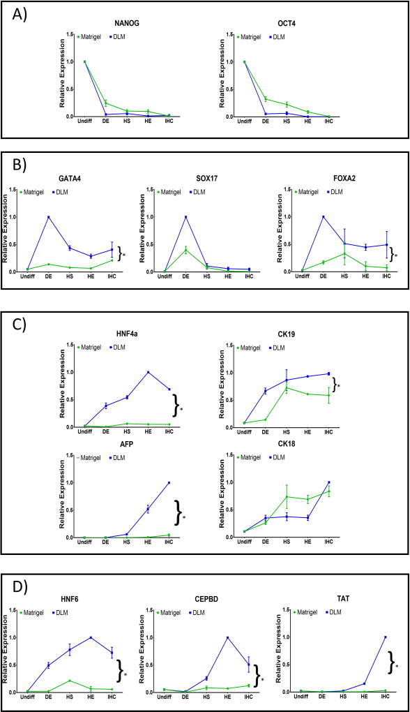
Figure 4. Expression of developmental stage-specific markers.
Cells differentiated on hDLM (blue lines) or Matrigel substrates (green lines) were analyzed at the end of each stage of the differentiation protocol for markers specific to (A) pluripotency (NANOG, OCT4), (B) definitive endoderm (GATA4, SOX17, FOXA2), (C) hepatoblast (HNF4α, CK19, CK18, AFP) and (D) mature hepatocytes (HNF6, CEBDP, TAT). (Results were normalized to the highest expression level of each gene achieved under all conditions)(n = 3, error bars represent standard deviation, *p<0.05).

