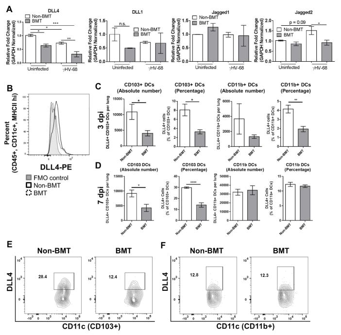
Figure 1. Loss of Notch ligand expression in lung associated APCs following BMT.
(A) Total lung CD11c+ cells were magnetically isolated (n = 4 mice per group) and infected ex-vivo with γHV-68. 24 hpi, indicated transcript expression level was analyzed via qRT-PCR. (B) Flow cytometric analysis of DLL4 expressing APCs. Cells were first gated on CD45+, CD11C+, and MHCII+ before gating on DLL4. Expression is compared to a flow minus one (FMO) control. (C and D) DLL4 expression in CD11c+ subsets were assessed by flow cytometry. Cells were first gated on CD45+, CD11c+, MHCII+, CD64− and then subdivided as either CD103+ or CD11b+. Graphed are absolute number or percentage of both CD103+ or CD11b+ DCs. Mean values are graphed (+/− SEM), statistical significance was calculated using ANOVA (panel A) or student’s T-test (panel c), * = p < 0.05, ** = p < 0.01, *** = p < 0.001, n.s. = not significant. (E and F) Representative flow plots showing percentages of DLL4+ cells from either CD103+ or CD11b+ DCs.

