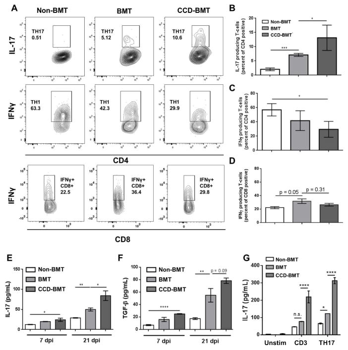
Figure 3. Loss of T-cell Notch signaling increases production of IL-17 from CD4+ lymphocytes following BMT.
Control or BMT mice (n = 4 – 5 mice per group) were infected with γHV-68 for 7 d. Lungs were harvested and collagenase digested. Following PMA and Ionomycin stimulation, cells were analyzed by flow cytometry for either IL-17 or IFNγ producing CD4+ or CD8+ T-cells. (A) representative flow-plots showing percentages of Th17, Th1, or IFNγ+ CD8+ cells present in the lungs of control, BMT or CCD-BMT mice at 7 dpi. (B, C, and D) Quantification of data represented in (A). (E and F) Leukocytes from collagenase digested lungs were purified by centrifugation through Percoll. Cells were cultured overnight in complete media, without additional stimulation, and clarified supernatants were analyzed by ELISA. (G) CD4+ T-cells were magnetically purified from spleens of either WT, BMT or CCD-BMT mice (n = 3 – 4 mice per group), 1x10⁶ cells were cultured in the presence of the indicated skewing condition for 24 h after which supernatants were harvested and analyzed for the indicated cytokine by ELISA (Unstim = no additives, CD3 = 1 μg/mL αCD3 and αCD28, Th17 = αCD3, αCD28, with 2 ng/mL TGFβ and 20 ng/mL IL-6). Statistical significance was calculated by ANOVA * = p < 0.05, ** = p < 0.01, *** = p < 0.001, **** = p < 0.0001.

