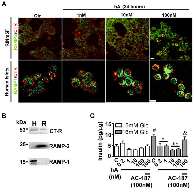
Figure 4. hA regulates trafficking of AM-R and insulin release in β-cells.
(A) Immunoconfocal microscopy analysis revealed expression and location of RAMP2 (green)/CT-R (red) in RINm5F β-cells (top panel) and RAMP1 (green)/CT-R (red) in human islet cells (bottom panel). Note increased recycling on AM-R components, CT-R and RAMPs, to the plasma membranes (yellow puncta) following exposure to increasing hA concentration (1–100 nM). Bar 10 μm. (B) Western blot analysis shows expression of CT-R and two RAMPs isoforms RAMP1 in human islets (H) and RAMP2 in RINm5F β-cells (R). (C) The inhibitory effect of hA on glucose-evoked insulin release from human islets was reversed by addition of AM-R antagonist, AC-187, indicating an AM-R mediated process. Intact human islets were exposed to normal (5mM) or high (16mM) glucose (Glc) concentrations in the presence or absence of hA (0.2–100 nM) and/or AC-187 (100 nM) for 30 minutes, following which insulin content in the samples (release) was analyzed by ELISA. Data was normalized to total protein content in samples. #p<0.05, 5mM Glc vs. 16mM Glc, n=6, **p<0.01, control vs. hA 0.2–100nM; and &p<0.05, hA 100nM vs. hA 100nM +AC-187 100nM, n=6. Significance established by ANOVA followed by Dunnett-Square test.

