Figure 1.
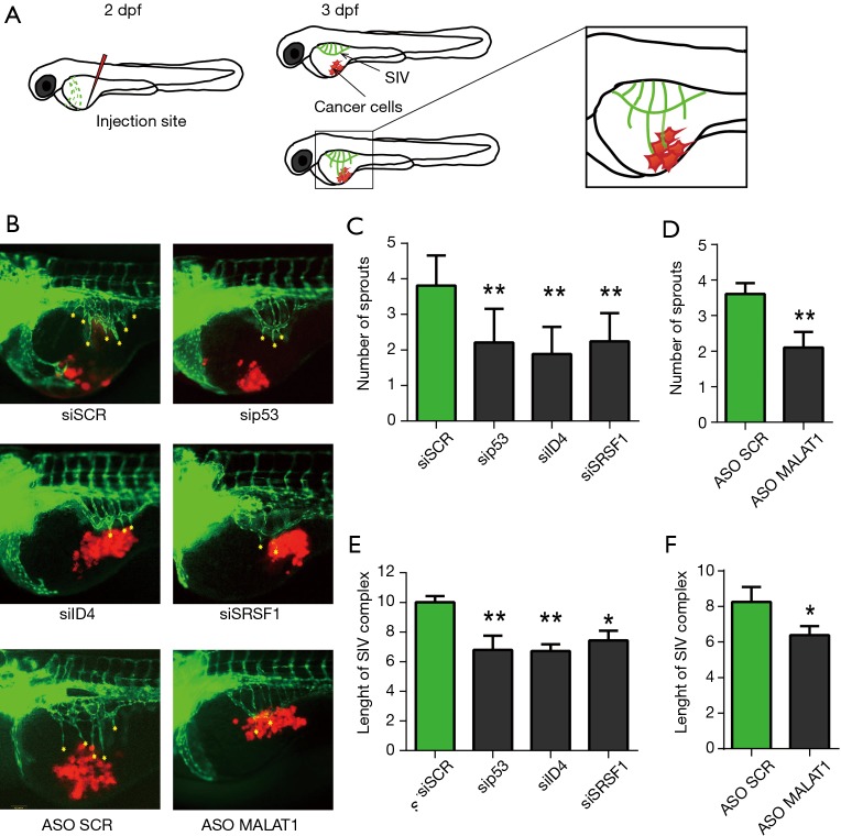
Silencing of p53, ID4, SRSF1, MALAT1 genes attenuates the proangiogenic properties of breast cancer cells in zebrafish embryos. (A) The scheme shows the injection site indicated by a red “needle”. Cancer cells (red) are injected into zebrafish embryo 2 days post fertilization (2 dpf). At this stage of development big blood vessel called duct of Couvier (green) is visible. At 3 dpf duct of Couvier disappears and a SIV complex is formed. Magnification shows a fragment of a fish presented in the pictures below; (B) representative pictures presenting SIV complex (green) growth in response to injection of cancer cells SKBR3 (red) depleted of p53, ID4, SRSF1 or MALAT1; (C-F) the graphs present a mean number of sprouts (C,D) and SIV complex length in arbitrary units (E,F) calculated from three independent experiments. In every experiment 20 embryos per condition were analyzed. *, P≤0.05; **, P≤0.01; one way ANOVA.

