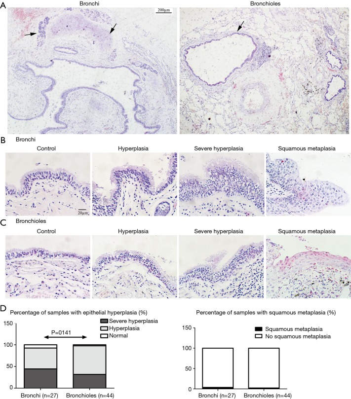
Figure 1.
Epithelium of bronchi and bronchioles. Bronchi has large lumen, pseudostratified columnar epithelium and hyaline cartilage plates with glands in the wall, which were showed by “↑”. Bronchioles simple cuboidal epithelium without cartilage and glands in the wall, and goblet cell disappear. “↑” showed an enlarged bronchioles (as compared to the adjacent pulmonary vein) (A, ×40 magnification; scale bar =200 µm). The common epithelium, epithelial hyperplasia, severe hyperplasia and squamous metaplasia in bronchi (B, ×400 magnification; scale bar =20 µm) and in bronchioles (C, ×400 magnification; scale bar =20 µm). Comparison of epithelial hyperplasia and squamous metaplasia (D, Fisher exact test) in bronchi and bronchioles.

