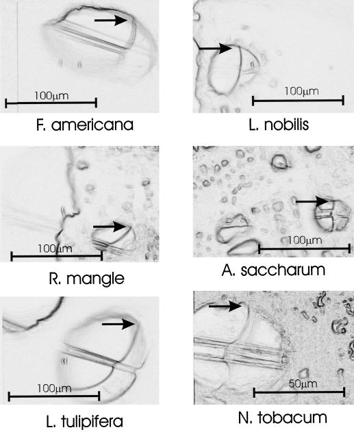
Figure 3.
Exemplar images (one per species) used in determining contact angle. Each frame shows the superposition of the vessel with water droplet in place over the same image after the droplet had been retracted into the capillary tube such that the outline of the vessel wall was more distinct. The focus was not altered between images. Arrows mark the point at which the contact angle was measured.

