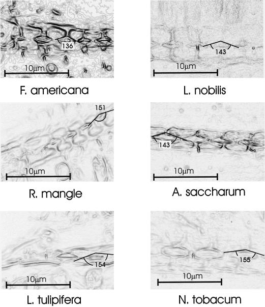
Figure 4.
Exemplar images (one per species) used in determining the angle of the pit chamber (2α). Freehand cross sections were photographed using a compound microscope (1,000× magnification, oil immersion). Image contrast was enhanced using the “find edges” feature of the image process program (Corel Photo-Paint), which in effect, transforms the image into a topographic map. Tangent lines were drawn along the walls of the pit chamber and the angle of their intersection measured.

