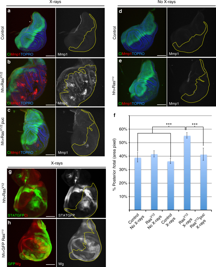
Fig. 5.
Constitutive expression of the Ras-MAPK pathway causes overgrowths due to permanent activity of the JNK pathway after irradiation. Images of wing discs of genotypes: a control tubGal80ts/+;hh-Gal4/+(n = 13), b UAS-RasV12/tubGal80ts;hh-Gal4/+(n = 9) and c tubGal80ts/UAS-RasV12;hhGal4/UAS-puc2A (n = 11) 72 h after irradiation. Non-irradiated wing discs of genotypes: d control tubGal80ts/+;hh-Gal4/+(n = 9) and e UAS-RasV12/tubGal80ts;hh-Gal4/+ (n = 10). Anterior compartments are stained with Ci (green). JNK activity is monitored by Mmp1 (red). Staining with TOPRO facilitates delimiting the discs. Note in b the large overgrowth of the posterior compartment that contains UAS-RasV12, associated with ectopic permanent JNK activity. Much of the overgrowth is prevented (c) when JNK function is compromised by overexpressing puc. There is no Mmp1activity (red) in non-irradiated discs (d, e). f Quantification of the posterior compartment area (in percentage) over the total disc area in the genotypes: non-irradiated tubGal80ts/+;hh-Gal4/+control (column: control No X-rays, n = 12), tubGal80ts/UAS-RasV12;hh-Gal4/+(column: RasV12 No X-rays, n = 6), and irradiated tubGal80ts/+;hh-Gal4/+control (column: control X-rays, n = 8), tubGal80ts/UAS-RasV12;hh-Gal4/+(column: RasV12 X-rays, n = 9) and tubGal80ts/UAS-RasV12;hhGal4/UAS-puc2A (column: RasV12 puc X-rays, n = 34). There is a statistically significant size increase of the posterior compartment after irradiation when RasV12 is expressed, compared with non-irradiated discs and with irradiated controls (***p-value < 0.0004, Mann–Whitney U-test). However, if JNK activity is suppressed by overexpressing puc, the size of the posterior compartment significantly decreases (***p-value < 0.0001, Mann–Whitney U-test). Columns in the graph represent mean ± s.d. Images of wing discs of genotypes: g STAT-GFP/tubGal80ts;UAS-RasV12/hh-Gal4 (n = 20) and h UAS-GFP,tubGal80ts/UAS-RasV12;hh-Gal4/+(n = 9) 72 h after irradiation; g shows the ectopic expression of STATGFP (green) only in the posterior compartment (delimited by the lack of Ci in red) where RasV12 is expressed; h shows the ectopic expression of Wg (red) only in the posterior compartment (green) where RasV12 is active. Scale bars are 100 μm

