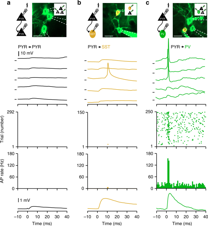
Fig. 2.
Single uEPSPs trigger action potentials in parvalbumin-expressing GABA-ergic neurons. a From top to bottom: in vivo Z-stack image and cartoon schematic showing experimental setup of whole-cell recordings from three PYR neurons, example Vm traces, action potential raster plot, peristimulus time histogram, and average unitary excitatory postsynaptic potential (uEPSP) in a connected, postsynaptic neuron in response to a single presynaptic AP in a neighboring PYR neuron (time of AP = 0 ms). APs were evoked during depolarized network activity (UPstate). b Same as a, but for an example SST neuron receiving an excitatory synaptic input from a neighboring PYR neuron. c Same as b, but for an example postsynaptic PV neuron. Note, uEPSPs trigger reliable and temporally precise APs in PV neurons. Scale bars, 20 µm. Vm marks in a–c show −50.0 mV

