Figure 5.
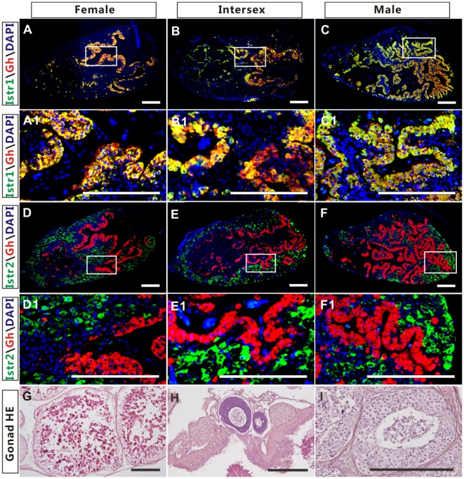
The co-localization of immunoreactive isotocin (Ist) receptor 1 (Istr1) (green) or Istr2 (green) with growth hormone (Gh) (red) in the pituitary of female, intersexual, and male ricefield eels. The rabbit antiserum against Istr1 (1:500) or Istr2 (1:500) and mouse antiserum against Gh (1:800) were used as primary antisera. The secondary antibodies were 1:500 diluted Alexa Fluor 488-labeled goat anti-rabbit immunoglobulin G (IgG) (H + L) for Istr1 and Istr2, and 1:500 diluted Cy3-labeled goat anti-mouse IgG (H + L) for Gh. DAPI was used to stain the nuclei blue. The images were observed and captured with a confocal microscope under the same conditions. (A1–F1) are higher magnification of (A–F), respectively. The overlapping of the red with the green color generated a yellow color. (G–I): HE-stained gonads of the experimental fish at female, intersexual, and male stages, respectively. Scale bar is 50 µm.

