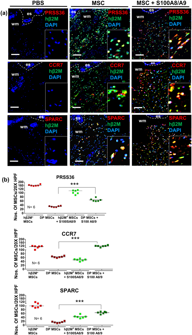
Figure 6.
Immunostaining for selected proteins encoded by up-regulated genes after injection of S100A8/A9 treated MSCs into wounds. Representative photomicrographs of sections from wounds which were injected with either control MSCs or S100A8/A9 treated MSCs. The expression of selected genes on protein level is shown with immunostaining for the serine protease PRSS36, the MSC specific chemokine receptor CCR7, and the developmental-like proteoglycan SPARC. Double staining (DP) for human β2 microglobulin (hβ2M is shown in green detecting the injected MSCs) and PRSS36 (shown in red) (a); CCR7 (shown in red) (b) and SPARC (shown in red) was compared to single staining with hβ2M. Nuclei were counterstained with DAPI. Scale bars: 200 μm. Dashed lines indicate the junction between epidermis (e) and dermis. es, eschar; wm, wound margin. Quantification of double-stained and single hβ2M+ MSCs in wounds injected with control MSCs and S100A8/A9 treated MSCs at days 2 after wounding were counted in 10 high-power fields per sample. Results are mean ± SD ratio of DP + and single hβ2M+ MSCs to total cells counted in the dermis (n = 6). ***P < 0.0001 versus non-treated control MSC injected wounds and MSCs pretreated with S100A8/A9 prior to be injected into wounds. The significance was obtained using ANOVA with Tukeys post hoc test.

