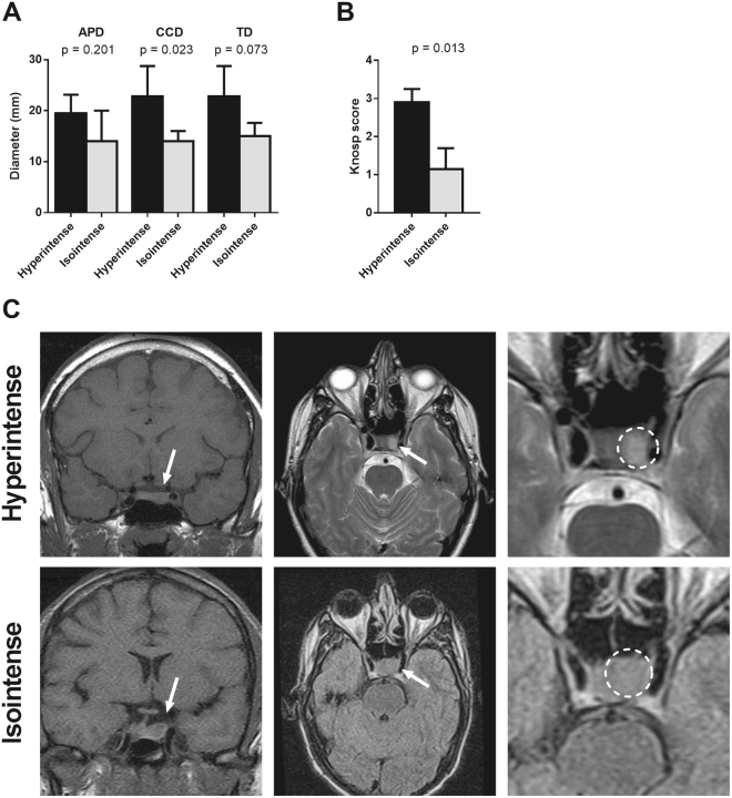
Figure 2.
Association between T2-signal and the diameter (A) and Knosp score (B) of somatotropinomas. The diameter of GH secreting adenomas was measured in millimetres. APD: anteroposterior diameter; TD: transverse diameter; CCD: craniocaudal diameter. Data represent median ± interquartile range. (C) Classification of T2-weighted signal of GH secreting pituitary adenomas: T2-hyperintense adenomas, upper-panel and T2-isointense adenomas, lower-panel. Tumour region was indicated with an arrow in coronal MRI (left), and in T2-weighted axial MRI (middle), the tumour area was indicated by a circle in right panel.

