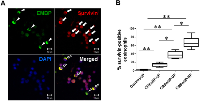
Figure 4.
Double immunofluorescence micrographs of EMBP and survivin expression in human NP tissue. (A) EMBP-positive cells (arrowheads) support survivin expression. Survivin protein (white arrows) was dominantly expressed in the cell cytoplasm, unlike in the nucleus. (B) Percentage of survivin-positive eosinophils (black arrows) was significantly greater in the NP tissue. Data are expressed as the means ± SD. *P < 0.05, **P < 0.01. Mann-Whitney U test. Scale bar = 10 μm.

