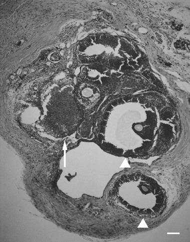
Figure 3.

An ovarian tissue specimen autotransplanted into the subcutaneous space. Corpora lutea (arrow) and growing follicles (arrow heads) including oocytes are seen. This ovarian section shows normal ovarian function although it was surrounded by fatty tissue. Bar, 100 µm.
