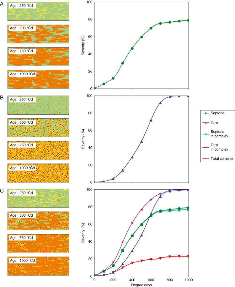
Fig. 4.
Growth of lesions of Z. tritici and P. triticina simulated with model 1. Left: pixels occupied by lesions. Right: severity curves over time expressed in degree-days (°Cd). Lines with symbols indicate the mean of 30 repetitions; lighter shades indicate the 95 % confidence interval. (A) Z. tritici alone. (B) P. triticina alone. (C) Z. tritici and P. triticina together.

