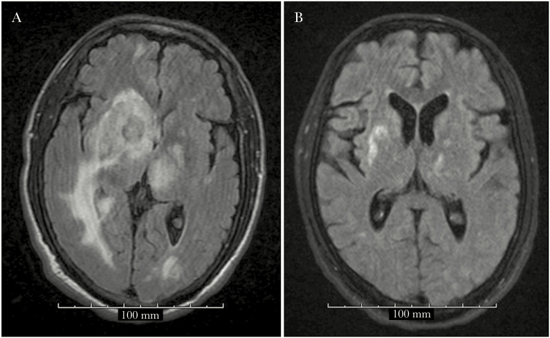
Figure 1.
Pre–pyrimethamine, leucovorin, and clindamycin (PLC) regimen: Magnetic resonance imaging (MRI) showing multiple bilateral T2 isodense round lesions with surrounding extensive edema and midline shift to the left (A). Post-PLC regimen: MRI showing interval decrease in lesion sizes and interval near complete resolution of the previously seen vasogenic edema (B).

