Figure 1.
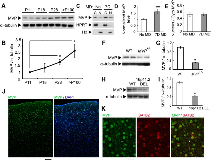
Expression of MVP proteins in cerebral cortex. A, B, Expression of MVP proteins during development and its quantification (normalized to P11; n = 3 animals for each time point). C, Expression of MVP in cytoplasm and nucleus after 7 d MD. HPRT and histone H3 served as cytosolic and nuclear markers, respectively. Lysates were collected from P35 animals. D, Quantification of MVP level after 7 d MD (n = 5 animals each). E, Ratio of nuclear/cytosolic MVP proteins after 7 d MD. F, G, Expression of MVP in WT and MVP+/− mice and its quantification (n = 3 animals each). Lysates were collected from P30 animals. H, I, Expression of MVP in WT and 16p11.2 microdeletion mice and its quantification (n = 3 animals each). Lysates were collected from P30 animals. J, MVP is expressed across all cortical layers. Scale bar, 100 μm. K, MVP is expressed in neurites (white asterisks) and nuclei. MVP is expressed in SATB2+ excitatory neurons (87 ± 2% colocalization, n = 3 animals). Scale bar, 25 μm. Averaged data are presented as mean ± SEM. *p < 0.05, **p < 0.01.

