Figure 4.
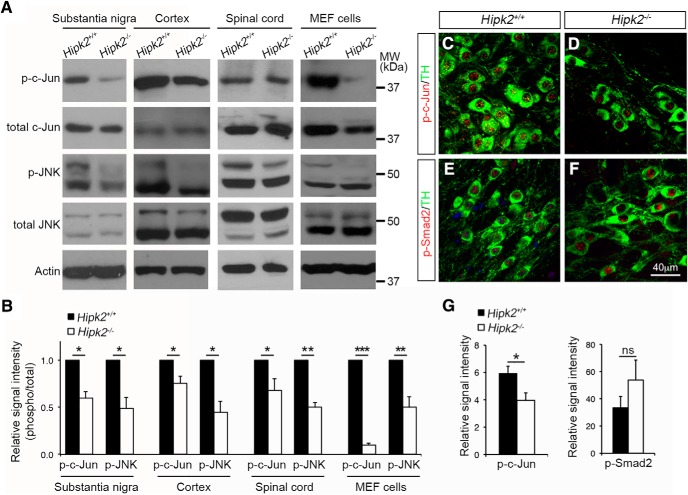
Reduced activation of the JNK–c-Jun signaling pathway in Hipk2−/− mouse brain. A, B, Western blots using lysates from the cerebral cortex, substantia nigra, and spinal cord of 2-month-old Hipk2+/+ and Hipk2−/− mice showed a consistent reduction in the levels of p-JNK and p-c-Jun, but no changes in total JNK and c-Jun. Similar results were identified in Western blots using protein lysates from Hipk2−/− MEF cells. The signal intensity of p-c-Jun, p-JNK, c-Jun, and JNK bands was quantified with National Institutes of Health ImageJ online software. C–F, Confocal microscopy of p-c-Jun and p-Smad2 expression in DA neurons of 2-month-old Hipk2+/+ and Hipk2−/− mice. G, Quantification of p-c-Jun and p-Smad2 fluorescent signal intensity in C–F. Data are mean ± SEM from at least three independent experiments. *p < 0.05 (ANOVA). **p < 0.01 (ANOVA). ***p < 0.001 (ANOVA). Not significant: p > 0.05.

