Figure 7.
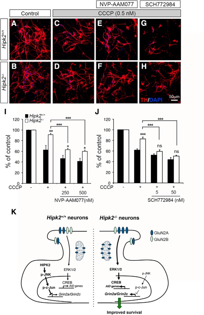
Pharmacological characterizations of CCCP-induced mitochondrial toxicity in Hipk2−/− DA neurons. A–J, Primary DA neurons from Hipk2−/− mice were more resistant to mitochondrial toxicity induced by CCCP than Hipk2+/+ DA neurons. CCCP toxicity in neurons was quantified by the percentage of surviving TH+ neurons based on confocal microscopy. Although blocking GluN2A with NVP-AAM077 increased the sensitivity of Hipk2−/− DA neurons to CCCP-mediated toxicity, the treated neurons were still more resistant to CCCP toxicity than Hipk2+/+ neurons. ERK inhibitor SCH772984 completely eliminated the resistance of Hipk2−/− DA neurons to CCCP-mediated toxicity. Data are mean ± SEM from at least three independent experiments. *p < 0.05 (ANOVA). **p < 0.01 (ANOVA). ***p < 0.001 (ANOVA). Not significant: p > 0.05. K, Schematic diagram depicting the working model by which the HIPK2-JNK pathway regulates c-Jun-mediated suppression of Grin2a and Grin2c in Hipk2+/+ neurons, which maintain the GluN2A/GluN2B ratio to regulate synaptic transmission and Ca2+-mediated ERK-CREB signaling pathway. Loss of HIPK2 or inhibition HIPK2 kinase activity by its specific inhibitors reduces JNK–c-Jun signaling, which derepresses the transcription of Grin2a and Grin2c, alters the GluN2A/GluN2B ratio, and activates ERK-CREB signal pathway that promotes the expression of AID genes to promote the survival of Hipk2−/− neurons in toxicity-induced by mitochondrial toxin CCCP.

