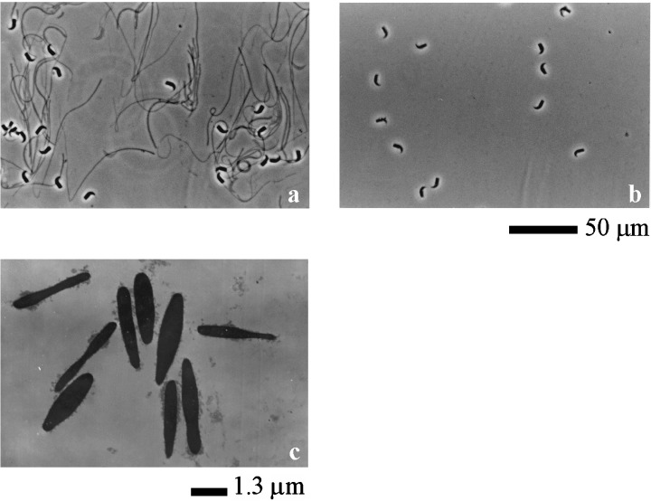
Figure 1.

Observation of extracted spermatozoa under microscope and electron microscope. (a) Spermatozoa which were extracted by the urea solution. Sperm head and fibrous sheath were observed under microscope. (b) Spermatozoa which were extracted by the urea‐thiourea solution after urea extracting. Only sperm head was observed under microscope. Scale bar shows 50 µm. (c) Spermatozoa which were extracted by the urea‐thiourea solution after urea extracting under electron microscope. Only nucleus of sperm head was observed under electron microscope. Scale bar shows 1.3 µm.
