
Figure 1.
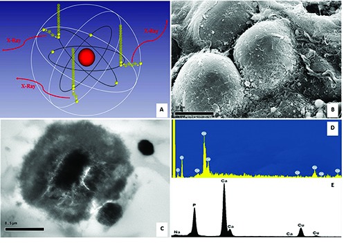
SEM and TEM EDX microanalysis. A) Characteristics X-rays was originated from the transitions of electrons from higher shells to lower shells by atoms of the specimen interacted with electrons beam. B) Scanning electron micrograph shows breast adipocyte cells (1000x). C) Transmission electron micrograph displays a psammomabody in a carotid plaque (30,000x). D) The Osmium used for lipid fixation is detected by SEM-EDX microanalysis. E) EDX spectrum demonstrate that psammoma-body is made of hydroxyapatite.