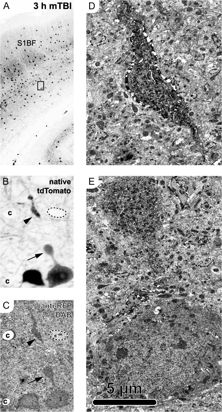
Figure 3.
Ultrastructural analysis of tdTomato+ PSAI identified via confocal microscopy. The same tdTomato+ profile was followed from the light (A,B) to electron (C–E) microscopy level at 3 h post-mTBI. (A, B) Confocal images capturing native tdTomato signal. (A) Overview image of S1BF with inset in layer 5/6 corresponding to tdTomato+ neuron in B, showing PSAI (arrow, oriented toward pia) and related disconnected distal segment (arrowhead). Employing the same RFP antibodies used for photostabilizing allowed use to follow the same tdTomato+ neuron from confocal (B) to electron (C–E) microscopy level. (C) The tdTomato+ neuron was identified based on morphology, including the perisomatic axonal swelling (arrow) and disconnected distal axonal segment (arrowhead), as well as other fiduciary markers including an adjacent tdTomato+ neuron (left), nearby capillaries (c), and a non-tdTomato expressing neuron adjacent to the tdTomato+ disconnected distal axonal segment (outline, upper right). (D–E) Ultrastructural analysis of the tdTomato+ distal disconnected segment (D) showed disorganized cytoskeleton consistent with the onset of Wallerian degeneration and the perisomatic swelling (E) laden with organelles and vesicles, indicating a focal site of impaired axonal transport.

