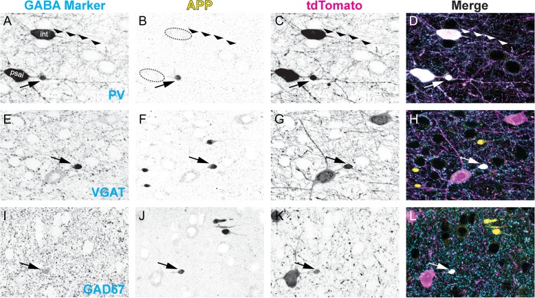
Figure 4.
GABAergic markers accumulate in APP+/tdTomato+ perisomatic axonal swellings. Representative images at 3 h post-mTBI showing GABAergic markers (A,E,I) with respect to APP immunoreactivity (B, F, J), which accumulates at focal sites of impaired axonal transport (arrows). Colocalization of tdTomato+ PSAI with PV (A–D), VGAT (E–H), and GAD67 (I–L) immunoreactivity confirms GABAergic interneuron axonal injury. (A) Normal uninjured intact (int) axonal profile (wide arrowheads) juxtaposed by PV+ interneuron PSAI (arrow). (B) APP is not detected within the intact axonal profile, while robust APP immunoreactivity colocalizes with tdTomato+/PV+ interneuron PSAI (C, D). Within sites of tdTomato+ PSAI (C,G,K), the immunoreactive profiles of VGAT (E) and GAD67 (I) are similar to APP+ axonal swellings (F and J, respectively). Qualitatively, the GAD67+ axonal swelling profile (I, L) has a better signal-to-noise than VGAT (E, H). Note non-GABAergic APP+ axonal swellings have opposite trajectories.

