
Fig. 2.
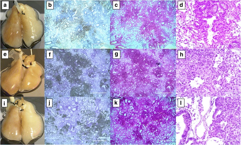
Non small cell lung cancer (NSCLC) cells on the cellular 4D lung model. Primary tumors were formed in a bronchocentric fashion on the cellular lung upon A549 (a–d), H1299 (e–h) and H460 (i–l) cell seeding though the trachea. Human mitochondrial IHC staining clearly shows the presence of a tumor formed by human lung cancer cells in a rat lung after 12 days of culture in a bioreactor. Low and high power H&E staining indicates the presence of a microscopic tumor with distinctive histology based on type of cell placed in the model. A549 cells formed a focal acinar pattern with enlarged nuclei and scattered atypical mitotic cells (c and d). H1299 cells formed a solid tumor and resemble poorly differentiated cells with scattered atypical mitosis (g and h). H460 cells formed a solid tumor with vague glandular formation (k and l)