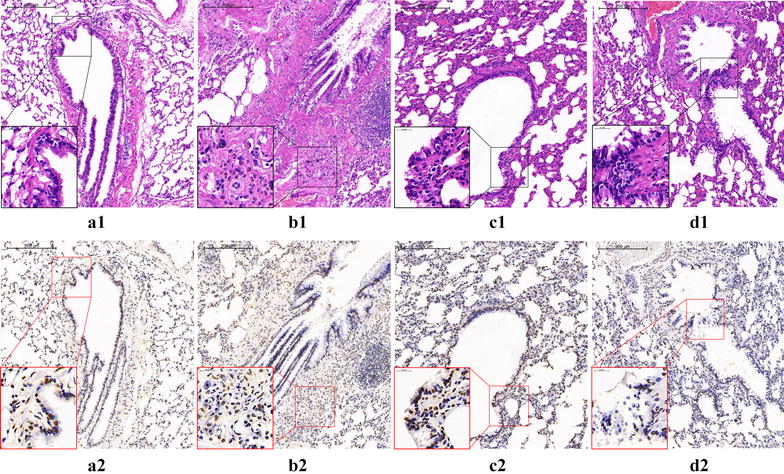
Fig. 6.

EOS apoptosis in lung tissue of rats: (a1, 2) Normal control group: The structure of bronchopulmonary tissue is normal, the lumen is smooth and the alveolar septum is intact. The eosinophil infiltration can be seen occasionally, and only a small amount of eosinophil apoptosis was found. (b1, 2) Model group: The bronchial lumen is stenotic, the epithelial cells proliferate and fall off, the tracheal smooth muscle and the alveolar septum thickens, and the structure is disordered. A large number of inflammatory cells, mainly eosinophils, infiltrating and apoptosis of eosinophils can be seen. (c1, 2) DXM group: Compared with the model group, the inflammatory cell infiltration of bronchopulmonary tissue in rats was alleviated, the pathological damage was significantly reduced, the apoptosis of EOS and the apoptotic index increased. (d1, 2) WTD group. Showed similar effects to DXM group in reducing pathological injury and increasing EOS apoptosis index in rats
