Fig. 6.

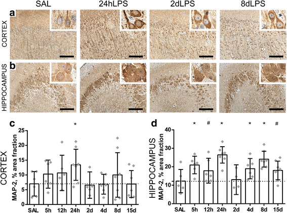
Intra-amniotic exposure to LPS results in altered dendritic development in the gray matter of the fetal brain. A significant increase of the area fraction (%) of MAP-2 immunoreactivity (IR) was found in the cerebral cortex at 24 h after LPS exposure compared to controls (SAL vs 24 h LPS p = 0.036) (a, c). In the hippocampus, an increase of area fraction (%) of MAP-2 IR was observed at 5, 12, and 24 h and at 4, 8, and 15 days after LPS exposure compared to controls (SAL vs 5 h LPS p = 0.007; SAL vs 12 h LPS p = 0.073; SAL vs 24 h LPS p = 0.000; SAL vs 4 days LPS p = 0.034; SAL vs 8 days LPS p = 0.000; SAL vs 15 days LPS p = 0.057) (b, d). Representative histological figures of MAP-2 in the cerebral cortex (a) and hippocampus (b) are depicted in control animals (SAL) and animals exposed to LPS for 24 h, 2 days, and 8 days. Images taken at × 100 magnification (insert at × 400 magnification), scale bar = 200 μm. Asterisk indicated p < 0.05 versus control (SAL); number sign indicated 0.05 < p < 0.1 versus control (SAL)
